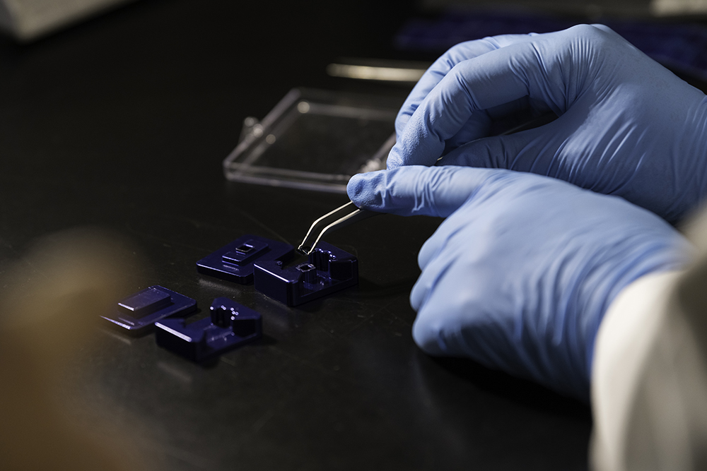
Biomedical Nanotechnology
What is Biomedical Nanotechnology?
Nanotechnology is developed by manipulating matter at 1 to 100 nanometers in size—bigger than what scientists normally consider molecules but smaller than bulk materials. As a result of their size, these nanomaterials have unique properties with a broad range of applications in medicine and basic biological research.
Areas of Focus
Nanomaterials have a broad range of applications in medicine and basic biological research. Some of those areas that are currently being explored by Rochester researchers inlucde:
Nanostructured Materials for Biological Sensing
Advancements in characterizing the genomes and proteomes of humans created new protein and nucleic acid targets for the biomedical research community to explore. Nanostructured materials can act as substrates for new biomedical sensors that power medical diagnostic systems.
Nanoporous Membranes
Rochester researchers developed novel silicon membranes with well-defined, tunable nanometer-sized pores that have great potential for improving the efficiency and speed of biofiltration.
Nanoparticle-Based Drug Delivery
Developing new polymer nanoparticles holds promise as a useful method for encapsulating therapeutics and delivering them effectively to the right target sites in the body.
Semiconductor Nanocrystals
Our researchers investigate imaging, transporting, and toxicity properties of semiconductor nanocrystals. “Quantum dots” are emerging as useful reagents for biomedical imaging, but little is known about the interactions of these materials with biological tissues, and their eventual fate in the body.
Nanobiomechanics
Researchers use the atomic force microscope, optical “tweezers,” magnetic tweezers, bioforce probes, and microcantilevers to study the mechanics that influence life at the sub-cellular level.