Medical Imaging

What is Medical Imaging?

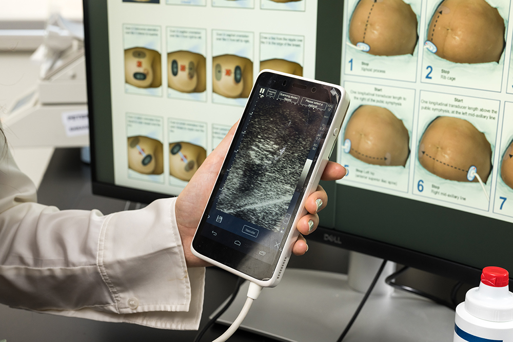
Medical imaging uses advanced technologies like X-rays, ultrasound, computerized tomography (CT) scans, and magnetic resonance imaging (MRI) to look inside the human body. These tools help doctors and technicians diagnose, monitor, and treat illnesses or injuries. The field involves developing the machines and the software that make these images possible, with the goal of improving healthcare and discovering new ways to treat patients.

Areas of Focus

Using these advanced imaging techniques, our biomedical engineers conduct cutting-edge research to:

  • Study bone health, joint health, infections, and treatments.
  • Investigate cancer biology, cancer pathology, and tumor development.
  • Predict flare-ups in autoimmune diseases and chronic illness management.
  • Develop injury biomechanics models and evaluate treatment options.
  • Fabricate and monitor 3D engineered tissues for biomedical applications.
  • Develop therapeutic applications, including drug delivery systems, wound healing solutions, thrombolysis, high-intensity focused ultrasound surgery, and gene transfection therapies.
  • Analyze complex medical imaging data for improved diagnostics and treatment planning.
A hand holding a device displaying an ultrasound image.

Revealing the Unseen

Medical Imaging at Rochester

Our department takes an interdisciplinary approach to medical imaging research, benefited greatly by our proximity to the University of Rochester Medical Centerand co-location with the Institute of Optics.

Medical imaging research here at Rochester covers a wide range of imaging methods and applications. We have continued to make meaningful advancements in the biomedical field, like discovering how ultrasound can be leveraged to build new blood vessels in living tissue.

Medical Imaging Researchers

Meet the faculty at the forefront of medical imaging research.

Interesting in doing research here as a graduate student? Check out our doctoral training faculty page for a list of biomedical engineering faculty who work with graduate students. 

Headshot of Timothy Baran.

Timothy Baran, PhD

Diffuse optics, photodynamic therapy, and medical image processing

Headshot of Edward Brown.

Edward Brown III, PhD

Multiphoton laser scanning microscopy, novel in vivo imaging and measurement techniques, tumor biology, angiogenesis

Headshot of Benjamín Castañeda.

Benjamín Castañeda, PhD

Development of Point-of-Care Technologies for resource limitted settings. Development of ultrasound-based imaging diagnostic techniques

Headshot of Regine Choe.

Regine Choe, PhD

Diffuse optics for in vivo cancer detection, diagnosis and therapy monitoring

Headshot of Diane Dalecki.

Diane Dalecki, PhD

Biomedical ultrasound, acoustics, lithotripsy, biological effects of ultrasound

Headshot of Marvin Doyley.

Marvin Doyley, PhD

Tissue characterization, inverse problems, breast imaging, elastography, cardiovascular disease, molecular imaging, ultrasound, and MRI

Headshot of Michael Giacomelli.

Michael Giacomelli, PhD

Multiphoton microscopy, surgical imaging, digital pathology, fluorescencelifetime imaging, 3D and molecular imaging

Headshot of Amy L. Lerner.

Amy L. Lerner, PhD

Orthopaedic biomechanics, bone growth and development, cartilage mechanics, medical image-based finite element modeling, knee biomechanics

Headshot of Susana Marcos.

Susana Marcos, PhD

Ocular Imaging, Corneal Biomechanics, Ocular implants, Opto-mechanical modeling in the eye, Myopia, Presbyopia, Cataract, Adaptive Optics, Visual Simulation, Diagnostic technologies in ophthalmology

Headshot of Stephen A. McAleavey.

Stephen A. McAleavey, PhD

Development of novel, clinically applicable ultrasound imaging techniques

Headshot of Mohammad Mehrmohammadi.

Mohammad Mehrmohammadi, PhD

Develop novel, hybrid, and ultrasound-based diagnostic methods, and define the clinical utility of the developed technologies as it applies to detection, diagnosis, and therapy of various pathologies

Headshot of Kevin James Parker.

Kevin James Parker, PhD

Medical imaging, digital imaging, halftoning, and novel scanning techniques using Doppler shift effects

Headshot of Jannick Rolland.

Jannick Rolland, PhD

Optical instrumentation, system engineering, optical coherence tomography

Headshot of Edward M. Schwarz.

Edward M. Schwarz, PhD

Pro-inflammatory cytokine signal transduction and novel drug and gene therapies for Rheumatoid Arthritis

Headshot of Axel W. E. Wismuller.

Axel W. E. Wismuller, MD, PhD

Intelligent image acquisition and analysis systems in biomedicine

Headshot of Chao Xie.

Chao Xie, PhD

Bone repair and regeneration for critical defects caused by trauma, infection, and tumors, aiming to connect translational medicine with innovative therapies for musculoskeletal diseases

Headshot of Jianhui Zhong.

Jianhui Zhong, PhD

Development and medical application of magnetic resonance imaging