Overview

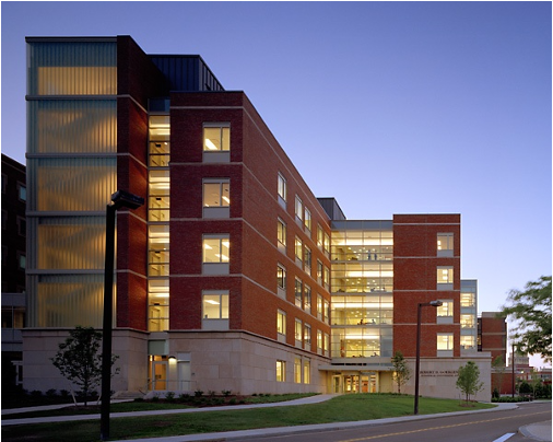
High Content Imaging Core (HCIC): The High Content Imaging Core is a 500 sq ft. facility in Goergen Hall that provides researchers at the University of Rochester access to high speed confocal and fluorescent microscopy. The core is partitioned into separate imaging compartments fitted with heavy durability blackout curtains to maintain fluorophore lifetime and limit errant light. The core has a a HERAcell VIOS 160i CO2 incubator for sample staging prior to imaging, dissection microscopes, benchtop refrigerator and freezer, and a wet bench for sample preparation.
Major imaging instruments include:
- Andor Dragonfly spinning disk confocal microscope on a Nikon Ti Eclipse Epifluorescent base with two Andor Sona sCMOS detectors, TIRF, and laser ablation/activation capabilities
- Incubator-based Etaluma LS850 Lumascope for long-term live sample imaging
- Leica SP5 Laser Scanning Confocal Microscope with environmental chamber for live-cell imaging
- Lumicks C-Trap optical tweezers system with two optical traps and automated microfluidics apparatus
- Nikon Ti-Eclipse 2 Epifluorescent microscope with Andor Zyla sCMOS and stage-based incubator for live cell and time-lapse microscopy
- Zeiss LSM980 with Airyscan 2 technology
- FIRE (Freeform Image Rotation to the Excitation plane) lightsheet microscope custom-built by a UR team spanning the biology, biomedical engineering, optics, and data science departments
Additionally, a dedicated workstation in the core contains a license for Imaris (Bitplane), an image analysis software with outstanding three-dimensional image analysis capabilities, and a Nikon Batch Deconvolution software package.
