ECE Seminar Lecture Series
From Vibrations to Innovations: The Biomedical Symphony of Nanobubbles
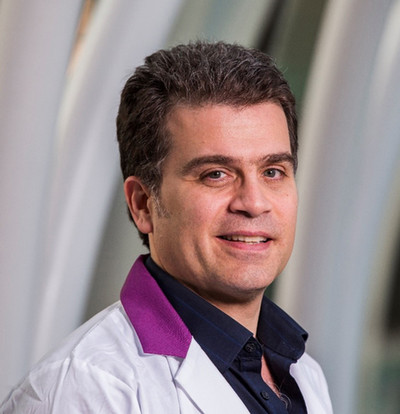
Michael C. Kolios, Professor, Toronto Metropolitan University
Wednesday, January 29, 2025
Noon–1 p.m.
601 Computer Studies Building

The most common imaging modality in the world is ultrasound (US) due to its affordability, safety, versatility, real-time imaging and ease of use. Contrast agents in ultrasound imaging improve the quality and interpretability of images by enhancing the visibility of specific structures or abnormalities. The most common contrast agents are microbubbles. Microbubbles are comparable to the size of a red blood cell and typically consist of a polymer or lipid shell encapsulating a gaseous core. However, micron-sized bubbles are intravascular agents and cannot probe the extravascular space to attach to cellular structures of interest. This limits the use of US molecular imaging to applications that involve microbubbles adhering to endothelial cells. Bubbles that can extravasate from the vasculature are needed to develop robust US molecular imaging applications. To achieve this extravasation, nanoscale contrast agents are required. Nanobubbles are sub-micron gas-filled structures, typically around 200 nanometers in diameter. While nanobubble contrast agents can escape the vasculature in principle, the resonant frequencies of nanobubbles are in order of 100s of MHz (for bubbles with a diameter of 200 nm or less). As the frequencies used in clinical imaging are far from the NB resonance, it was thought that decreasing bubble size to the nanometer scale would lead to resonant frequencies for which there would be no detectable signal at the clinically relevant US frequencies (~ 10MHz or lower).
Recent work with long-circulating nanobubble contrast agents has shown that contrast-enhanced ultrasound (CEUS) approaches based on non-linear imaging schemes can achieve high contrast. We have hypothesized that the key to the NB's effectiveness as a contrast agent are the non-linear properties of the stabilizing shell as the bubble oscillator interacts with the incident ultrasound waves. In this presentation, we will explore the physics of the complex response of the bubble oscillator to ultrasound using bifurcation diagrams, show how the shell properties impart strong non-linearities to the bubble oscillator, and how bubbles interact with each other. Through in-vitro and in-vivo experiments, we will discuss the mechanisms underlying nanobubble behavior in biological systems, showcase the evidence that NBs extravasate, and discuss the current challenges in translating these technologies from bench to bedside. By showcasing new nanobubble imaging applications, this presentation aims to provide insights into how nanobubbles may shape the future of US molecular imaging.
Dr. Michael C. Kolios is a Professor in the Department of Physics at Toronto Metropolitan University and Associate Dean of Research, Innovation and External Partnerships in the Faculty of Science. His work focuses on using ultrasound and optics in the biomedical sciences. Dr. Kolios leads a large group focusing on optical and ultrasound methods to characterize tissues and disease and develop theranostic agents that will assist therapeutic and diagnostic applications. He has published over 200 peer-reviewed journal publications and five book chapters. Dr. Kolios has received numerous teaching and research awards, including the Canada Research Chair in Biomedical Applications of Ultrasound and the Ontario Premiers Research Excellence Award. In 2016, he received the American Institute of Ultrasound in Medicine (AIUM) Joseph H. Holmes Basic Science Pioneer Award for significant contributions to the growth and development of medical ultrasound. In 2023, he received the IEEE Carl Hellmuth Hertz Ultrasonics Award that recognizes investigators for their outstanding mid-career achievements and for promoting the field of Ultrasonics. He is on the editorial board of the journal Ultrasound Imaging and Photoacoustics and is a member of many national and international committees. He was a charter member of the National Institutes of Health (NIH) Biomedical Imaging Technology A study section and a member of the College of Reviewers for the Canadian Institutes of Health Research (CIHR).

