Three-dimensional sonoelastography imaging of HIFU-induced lesions in bovine livers
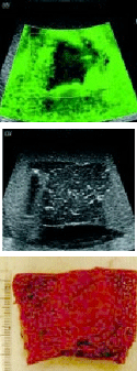
 High intensity focused ultrasound (HIFU) is a noninvasive surgical tool which produces tissue coagulation necrosis for killing the malignant tumor within a well defined volume in the tissue. As a real time lesion monitoring method, sonoelastography was investigated for the visualization ofHIFU-induced lesions in the bovine liver in vitro. Tissue samples (4x4x4 cm3) were cut from fresh bovine liver and then degassed overnight. Lesions were created in tissue samples by a HIFU transducer. The compounding lesion size was determined by the number of individual lesions treated with identical HIFU exposure dose. Three-dimensional sonoelastography images were acquired from the liverembedded agar phantom. Each lesion is displayed as a dark deficit area surrounded by a bright green background in the image. After imaging, lesions were examined by gross pathology to verify their size, shape and volume.
The gross pathology results showed that HIFU-induced compounding lesions were relatively uniform, palpably harder and brighter than the normal tissue and almost ellipsoidal in shape. The mean volumes of the three 2x2 compounding lesions and the two 3x3 lesions measured by fluid displacement were 2.3 cm3 and 6.2 cm3 , respectively. As the smallest lesion in the test, a 1x2 lesion with 1.5 cm3 in volume was also successfully detected by 3D sonoelastography imaging. In the sonoelastography images, the edge of each lesion was a little ambiguous in a range of ˜3 mm. When only the darkest region in the image was delineated as the lesion, the mean sonoelastography volume of the 6 lesions was 83% of the volume measured by fluid displacement. Good correlation between the lesion dimensions determined from sonoelastography images and gross pathology was also found. This study demonstrates that sonoelastography is a potential real-time method to accurately monitor the HIFU therapy of cancerous lesions.
Researcher: Kevin James Parker, Ph.D.
Medical imaging, digital imaging, halftoning, and novel scanning techniques using Doppler shift effects
