
Summer REU
On Hiatus for summer 2025. Check back for details about summer 2026!
National Science Foundation Research Experience for Undergraduates (NSF REU)
Can functional ultrasound detect neural activity with better sensitivity than functional magnetic resonance imaging (fMRI)? How well can diffuse optical techniques detect breast cancer and monitor bone healing? Does combining machine learning with ultrasound allow better flow detection in smaller vessels or increase the speed of processing large echo datasets (gigabyte-sized)? Can cell imaging methods help researchers determine the mechanisms by which COVID-19 and SARS-CoV-2 induce cellular stress? Can GSP-based graph learning frameworks be used to estimate structural brain connectivity from functional signals measured by resting-state fMRI?
Diverse teams are needed to answer engineering questions like these. This Imaging in Medicine and Biology REU site will provide an environment for undergraduates to complete individual research projects under the mentorship of leaders in their respective fields, in a world-class interdisciplinary research environment. The Imaging in Medicine and Biology REU site will also provide structured enrichment programs to participants that will allow students to develop critical skills needed to be successful in STEM graduate education (GRE preparation, time management, oral/written presentation). The University of Rochester (UR) Hajim School of Engineering and University of Rochester Medical Center (URMC) is an ideal setting for this REU site; it is the home of the Rochester Center for Biomedical Ultrasound (RCBU), the Institute of Optics, the Goergen Institute for Data Science and Artificial Intelligence (GIDS-AI), and the Del Monte Institute of Neuroscience—members of which will serve as mentors in this REU site.
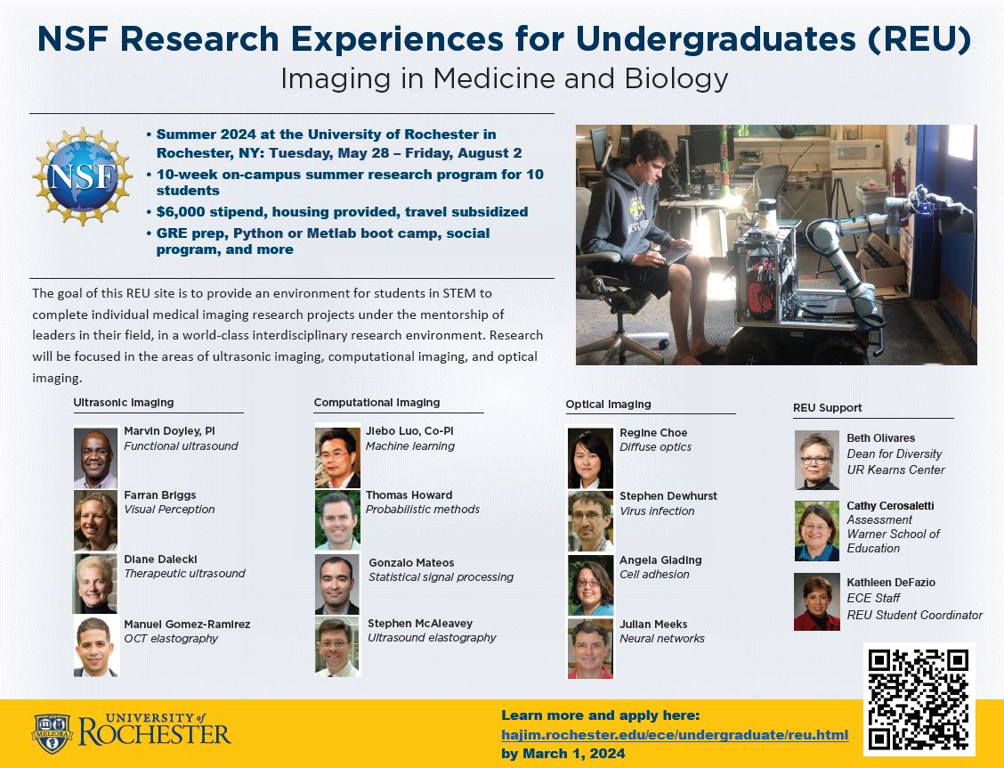
See the full abstract for this NSF REU for more information.
More information regarding the NSF REU program in general can be found on the full abstract for this NSF REU for more information.
Principal Investigator
Marvin Doyley, PhD
Professor and Chair
Department of Electrical and Computer Engineering
Co-Principal Investigator
Jiebo Luo, PhD
Professor
Department of Computer Science
How to Apply/Eligibility
You are eligible to apply if:
- You are a 1st, 2nd, or 3rd year full-time student at a college or university.
- You are a U.S. citizen or hold a green card as a permanent resident.
- You have little or no research experience.
We are unable to support international students via this federally-funded NSF REU program. If you are looking for other research opportunities at the University of Rochester, please visit the Office of Undergraduate Research. Alternatively, for self-funded research opportunities, you can reach out to one of the faculty members affiliated with this REU (see projects tab below) or reach out to any of the faculty of the Department of Electrical and Computer Engineering directly to discuss your research interests.
It is expected that all students accepted into this REU program will be fully vaccinated for COVID-19. Please visit the University of Rochester’s COVID-19 Resource Center for more information, including the current face mask guidelines.
Before starting the application, you should prepare:
- An unofficial college transcript, that is, a list of your college courses and grades, including the courses you are currently taking.
- Your CV or resume
- A 500-word essay explaining why you wish to participate in this REU, including how it engages your interests, how the experience would support or help you define your career goals, and special skills and interests you would bring to the program.
- The name and email address of a faculty member, instructor, supervisor, advisor, or mentor who can recommend you for the REU.
You can apply to this REU site using the NSF ETAP common application portal. Applications will be accepted through March 1, 2024. Notification of acceptance will be communicated between March 10 and April 1, 2024.
The REU Experience
The 2024 session for this REU site will take place from Tuesday, May 28 through Friday, August 2.
Students accepted into the REU will receive:
- On-campus housing
- Meal stipend
- Stipend of $6000 for other expenses and to take back home
- Up to $450 to help pay for travel to and from Rochester
Your experience will include:
- A MATLAB programming bootcamp that introduces students to image processing techniques such as image segmentation, principal component analysis, and multidimensional filter design, which are fundamental to medical imaging.
- Working with a team of students and faculty on one of the REU projects.
- Workshops on topics such as career planning and preparing for graduate school.
- Social events.
Summer activities for all REUs across the University of Rochester campus are organized by the David T. Kearns Center, including the REU orientation and undergraduate research symposium.
If you have any questions about the REU or the application process that are not answered here, please send an email to ece-reu@rochester.edu.
Projects
You will be able to specify your top project preferences from the list below. We will do our best to match you with one that matches your preferences and interests. You will be assigned to a project based on your background and skills.
2024 Projects (Planned)
Project #1
Title: High-resolution functional ultrasound imaging based on ultrafast Doppler ultrasound
Mentor: Farran Briggs (Neuroscience)
High-resolution functional ultrasound imaging (fUS) is an innovative method to gather large neuron activity data sets throughout the brain of rodents, ferrets, and non-human primates. Students will use fUS to visualize neuronal activity in visual brain areas. They will explore the impact of cortical feedback to the visual thalamus and its functional pattern of activation. At the end of the 10-week research experience, students should be able to (a) use fUS to image the visual cortex and visual thalamus, (b) perform basic histological processing, and (c) compute power Doppler images from ultra-fast plane-wave images.
Project #2
Title: Diffuse optical imaging for non-invasive deep tissue monitoring of bone healing
Mentor: Regine Choe (Biomedical Engineering)
Diffuse optical imaging can quantify hemodynamic parameters of deep-tissue in vivo samples non-invasively, enabling longitudinal monitoring of the bone graft vascularization process. The project will focus on developing and validating diffuse optical imaging methods. At the end of the 10-week research experience, students should be able to (a) perform 3D image reconstruction based on diffuse optical measurements, and (b) develop image processing methods to compare diffuse optical images and clinical images obtained using micro-CT, MRI, and ultrasound.
Project #3
Title: Advancing high-frequency ultrasound imaging technologies to visualize and quantitatively monitor structural changes in tissue
Mentor: Diane Dalecki (Biomedical Engineering)
In this project, students will develop quantitative ultrasound imaging metrics to characterize collagen microstructure within tendon and use machine learning techniques to test these quantitative metrics as noninvasive indicators of disease or injury. At the end of the 10-week research experience, students should be able to apply (a) quantitative, high-frequency ultrasound imaging techniques to monitor cell migration or proliferation, and (b) machine learning techniques to quantitative ultrasound data.
Project #4
Title: Imaging of virally induced exodosis
Mentor: Stephen Dewhurst (Microbiology and Immunology)
Students will use cell imaging methods to examine fundamental mechanisms by which human coronaviruses and SARS-CoV-2 induce cellular stress, and “ER exodosis” (i.e., the release of the protein contents of the endoplasmic reticulum from the cell). At the end of the 10-week research experience, students should be able to (a) determine whether the SARS-CoV-2 homologs of the genes shown to induce ER exodosis by SARS-CoV-1 (the “viroporins” E and 3a) are also able to induce ER exodosis, and (b) know how to examine protein assays from minimally pathogenic human coronaviruses.
Project #5
Title: Functional ultrasound brain imaging via neurovascular and neuromechanical coupling
Mentor: Marvin Doyley (Electrical and Computer Engineering)
In this project the student will perform studies using high-frequency functional ultrasound (fUS) and high-frequency shear wave elastography (SWE) in the developing brains of murine models. Students will compute activation maps from fUS and SWE images obtained with auditory stimulation. Studies will also investigate if advanced machine learning techniques provide better activation maps and allow the detection of smaller vessels. At the end of the 10-week research experience, students should be able to perform ultrasound imaging, compute activation maps from ultrasound SWE and power Doppler data, and construct activation maps with different approaches including machine learning methods.
Project #6
Title: Super-resolution imaging of vascular malformation development
Mentor: Angela Glading (Pharmacology and Physiology)
This project would require training in advanced optical imaging, and development of an analysis program to quantify protein co-localization. Students will perform transfection, confocal ratio-metric imaging (FRET or FLIM), and permeability measurements on primary endothelial cell cultures. At the end of the 10-week research experience, students should be able to perform staining, image acquisition, and contribute to analysis and subsequent creation of 3D models of the vascular tissue.
Project #7
Title: Bioluminescence to imaging of tactile sensory perception
Mentor: Manuel Gomez-Ramirez (Brain and Cognitive Science)
In this project the students will use bioluminescence to study the role of excitatory vs. inhibitory cells in tactile sensory perception. At the end of the 10-week research experience, students should be able to perform bioluminescence imaging, headpost implant, and viral injection.
Project #8
Title: Robot-assisted strain imaging
Mentor: Thomas Howard (Electrical and Computer Engineering)
In this project students will develop perception algorithms for robotically-assisted strain elastography, which uses a robotic arm to capture force and ultrasound measurements that are used to infer the stiffness and structure of underlying tissue. Students will use tissue models to evaluate the accuracy and quality of the resulting images and explore different organs such as the liver. Students will also learn software development and best practices for robotic systems. At the end of the 10-week research experience students should be able to calculate stiffness maps using strain imaging techniques with a robotic manipulator.
Project #9
Title: Determination of 3-D scapular position from plain radiographs using computer vision
Mentor: Jiebo Luo (Computer Science)
Abnormalities in scapular mechanics, or scapular dyskinesis, during shoulder motion negatively affects function. Diagnosis of scapular dyskinesis is important to guide treatment and improve patient outcomes but can be challenging to identify clinically. In this project, students will develop a computer vision algorithm to infer 3-D scapular position through interpretation of shoulder plain 2D radiographs. At the end of the 10-week research experience, students should be able to develop 2D and 3D landmark detection and registration methods and have a working knowledge of various registration methods such as RANSAC and mutual information.
Project #10
Title: GSP-based graph learning framework to estimate structural brain connectivity
Mentor: Gonzalo Mateos (Electrical and Computer Engineering)
In this project students will develop a GSP-based graph learning framework to estimate structural brain connectivity from functional signals measured by resting-state functional magnetic resonance imaging (fMRI). At the end of the 10-week research experience, students should be able to construct structural graph models of neural pathways in white matter and use statistical techniques to construct function graphs.
Project #11
Title: Ultrasound stiffness imaging using deep learning
Mentor: Stephen McAleavey (Biomedical Engineering)
In this project, the student will investigate the feasibility of using deep-learning algorithms to obtain detailed images of tissue mechanical properties (e.g., viscoelasticity, anisotropy). Students will also study the correlation between mechanical property images and disease/injury. At the end of the 10-week research experience, students should be able to apply machine learning methods to train a network to estimate tissue mechanical properties from shear wave data.
Project #12
Title: Big data imaging
Mentor: Justin Meeks (Neuroscience)
In this project students will analyze large imaging datasets obtained from LSM and 2-photon microscopes. Students will use these data sets to investigate neural networks and pathways that underlie mammalian social behavior. At the end of the 10-week research experience, students should know the theory and practice of producing, databasing, and analyzing large imaging datasets through LSM and 2-photon microscopy.